
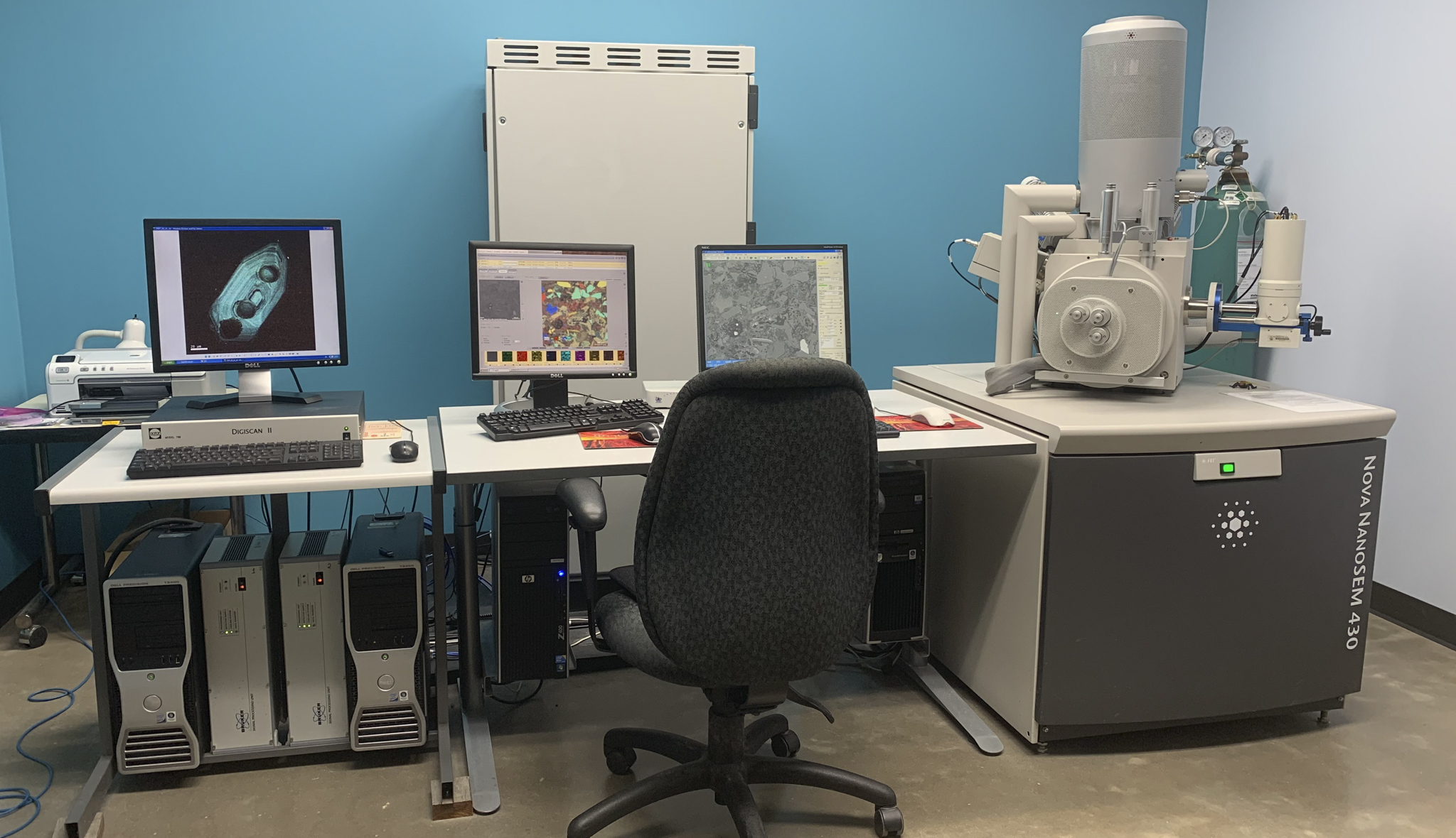
The FEI Nano Nova 430 FEG-SEM installed in 2009 is configured for high-resolution and analytical imaging of various materials. The instrument is equipped with a field emission electron source, oil-free vacuum system, Secondary Electron (SE), Back Scattered Electron (BSE), Energy Dispersive X-Ray Spectroscopy (EDS), magnetic immersion lens, low vacuum detector and chromatic Cathodoluminescence (CL) detector. Through-the-lens “immersion mode” imaging allows resolution of 3-4 nm under optimal conditions. The system consists of a large chamber with a 5-axes cartesian stage that can accommodate large samples. The low vacuum detector and helix detector enable to acquire images on uncoated rock samples. The Gatan chroma CL provides colored CL images to understand diagenesis in the rock samples.
Specifications:
- Spatial resolution: 3-4 nm under optimal conditions
- Accelerating Voltages from 0.5 kV - 30 kV
- Everhart Thornley Detector (for SE)
- Solid-state BSED for high vacuum
- In Lens SE (TLD SE) and BSE (TLD BSE) detectors
- Gatan Chroma CL Detector for wavelength 400-800 nm
- EDS X-ray mapping (dual Bruker 30 mm2 detectors)
- Low Vacuum Capability (< 0.1 Torr) using UHR low vacuum SED (Helix Detector) and low vacuum SED (LVD)
- Deceleration Mode, which allows to work at accelerating voltage of 2kV
- IR-CCD camera for in-chamber viewing
- 5-Axes motorized Stage (X-Y-Z-Tilt-Rotation)
- Working distance 5 mm -13 mm
- Beam current of up to 10 nA in High-Resolution Mode and up to 22 nA in Analytical Mode
Imaging modes:
Secondary electron (SE) Detector
Secondary electrons vary in intensity largely in concert with surface tilt and thus provide a topographic image. SE imaging can be carried out at very low beam energies and with an extremely small diameter beam, allowing for high spatial resolutions. This imaging mode is used to examine the smallest pores in mudrocks.
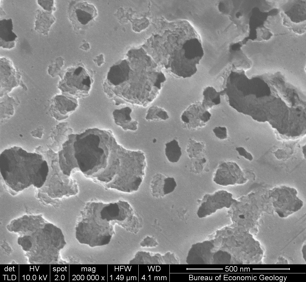
SE imaging allows close inspection of pores at the nanoscale. Such imaging has shown that many gas shales have pore systems that are located within organic matter. This fast opens the possibility of porosity prediction using an understanding of thermal maturation—a topic that is attracting much current investigation.
Back-scattered electron (BSE) Detector
BSEs carry information on composition. Although BSE images are generally produced at higher beam energies (and lower magnifications) than SE images, they yield information on rock fabric with immensely greater clarity than that of light microscopy.
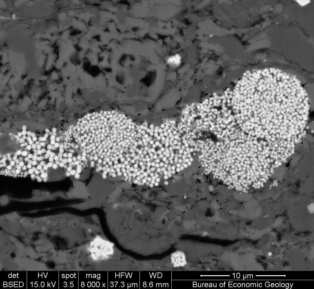
BSE imaging carries information that relates to a very thin slice of the rock. In an Eagle Ford shale sample, BSE reveals the sizes and shapes of pyrite crystals, fossil debris and organic matter with great clarity.
Cathodoluminescence (CL) Detector
The CL signal can be used to image variations in trace elements and crystal defects that are not readily visible in any other form of imaging. CL is especially useful for discriminating grains from cements in both silicate and carbonate systems.
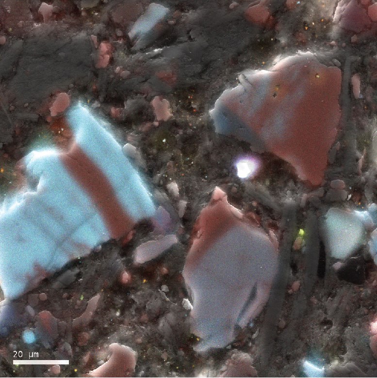
CL imaging in mudrocks can reveal microscale cementation that is an important control on mechanical rock properties.
X-ray energy-dispersive spectroscopy (EDS) Detector
X-ray signals go beyond BSE imaging to directly display mineral distributions. Mineral species depicted in false color can be readily interpreted as to component type. Use of a field-emission beam, combined with the approach of summing signals from multiple detectors, allows X-ray mapping at some of the highest resolutions possible.
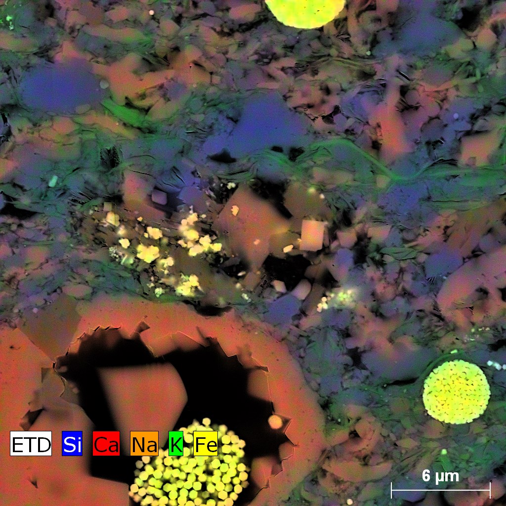
X-ray maps of Eagle Ford Shale. The complex composition of the silt and clay-size grain assemblage. Low left: a foraminifera chamber filled with calcite cement, pyrite framboid and migrated bitumen.
For training and using this tool, please contact Priyanka Periwal.
For inquiries on the instrumentation and capabilities please contact Rob Reed.

