
Hitachi SU-8700 FEG-SEM
The Hitachi SU-8700 FEG-SEM installed in 2024 brings in a high-resolution imaging on both coated and uncoated samples, analytical imaging, automated minerology, and large area mapping of various materials. The instrument is equipped with a Schottky field emission electron source with high probe current, oil-free vacuum system, three detectors (UD, MD and LD) in the column for Secondary Electron (SE) and Back Scattered Electron (BSE), insertable Photo-diode Backscattered electron (PD-BSE), Energy Dispersive X-Ray Spectroscopy (EDS), Variable pressure secondary electron detector (VP-SE) and chromatic Cathodoluminescence (CL) detector. The automated alignment of the electron optics enables stable setting of optical conditions. The ability of simultaneous capture of different channels (up to 6 channels) makes it an efficient system. The cold FEG with high probe current allows imaging at accelerating voltages as low as 500 V. The UD detector in the column allows resolution of ~1 nm under optimal conditions. The system consists of a large chamber with a 5-axis cartesian stage that can accommodate large samples. The Delmic Sparc Compact CL provides colored CL images to understand diagenesis in the rock samples.
Specifications:
- Spatial resolution: 0.8-1 nm under optimal conditions
- Accelerating Voltages from 0.5 kV - 30 kV
- Everhart Thornley Detector (LD for SE) and insertable photo-diode BSE Detector (PD-BSE for BSE) under high vacuum
- In column SE (UD) and BSE (MD) detectors
- Delmic SPARC Compact Detector for wavelength 400-800 nm
- Bruker EDS X-ray mapping (dual XFlash 7 detectors) with optimum geometry for efficient collection of the generated X-rays.
- Variable Vacuum ranging from 5-300 Pa
- Deceleration Mode, which allows to work at accelerating voltage of 2kV
- IR-CCD camera for in-chamber viewing
- 5-Axis motorized Stage (X-Y-Z-Tilt-Rotation)
- Working distance 1.2 mm – 15 mm
- Beam current of up to 200 nA in High-Resolution Mode and up to 2 nA in Analytical Mode
- Large area mapping for all detectors
- Mineral composition maps obtained by AMICS
Sample types:
Depending on the research question to be addressed, we work on different types of samples.
Imaging modes:
Upper detector: Secondary electron (UD) Detector
Secondary electrons vary in intensity largely in concert with surface tilt and thus provide a topographic image. SE imaging can be carried out at very low beam energies and with an extremely small diameter beam, allowing for high spatial resolutions. This imaging mode is used to examine the smallest pores in mudrocks.
SE imaging allows close inspection of pores at the nanoscale. Such imaging has shown that many gas shales have pore systems that are located within organic matter. This fast opens the possibility of porosity prediction using an understanding of thermal maturation—a topic that is attracting much current investigation.
Photo Diode Backscattered Electron (PD-BSE) Detector
BSEs carry information on composition. Although BSE images are generally produced at higher beam energies (and lower magnifications) than SE images, they yield information on rock fabric with immensely greater clarity than that of light microscopy.
.jpg)
BSE imaging carries information that relates to a very thin slice of the rock. In this Haynesville shale sample, BSE reveals the sizes and shapes of quartz and feldspar grains, pyrites, types of clay and organic matter with great clarity.
Cathodoluminescence (CL) Detector
The CL signal can be used to image variations in trace elements and crystal defects that are not readily visible in any other form of imaging. CL is especially useful for discriminating grains from cements in both silicate and carbonate systems.
CL imaging in mudrocks can reveal microscale cementation and quartz overgrowth that is an important control on mechanical rock properties.
X-ray energy-dispersive spectroscopy (EDS) Detector
X-ray signals go beyond BSE imaging to directly display mineral distributions. Mineral species depicted in false color can be readily interpreted as to component type. Use of a field-emission beam, combined with the approach of summing signals from multiple detectors, allows X-ray mapping at some of the highest resolutions possible.
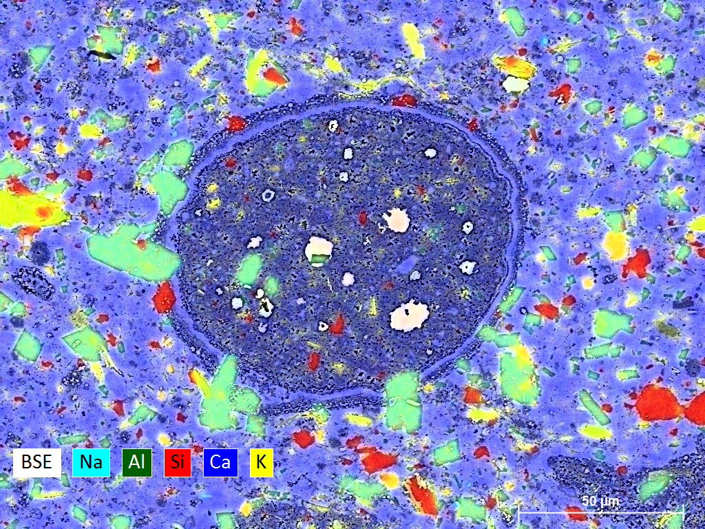
X-ray maps of Lacustrine Shale. The complex composition of the quartz, feldspars, clay-size grain assemblage. Center: algal cyst chamber filled with dolomite.
Large-area mapping
Automatic acquisition of images to obtain large area map with any detector of choice is available. A collection of tiles is collected and stitched to form a large area map.

Large Area X-Ray analysis on CM Murchison Meteorite, USNM-5487 taken on loan from Smithsonian Museum given to Dr. Romy Hanna. The matrix is comprised of Olivine, Diopside, Tochillionite, Chlorite.
Advanced Mineral Identification and Characterization System (AMICS)
For training and using this tool, please contact Priyanka Periwal.
For inquiries on the instrumentation and capabilities please contact Lucy Tingwei Ko.

